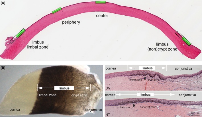
Figure 1.

Morphology of the equine cornea: overview of an entire cornea cross section with the four examined zones marked in green (A). Picture of a whole mount sample of the transitional zone from cornea to conjunctiva, clearly depicting two distinct subzones in the limbus and cross sections of these zones, showing crypts in dorso‐ventral (DV) sections and noncrypt zone in naso‐temporal (NT) sections (B). Note differences in the composition of the underlying stroma including vasculature which is distinctive in the different zones. Scale bar =100 µm
