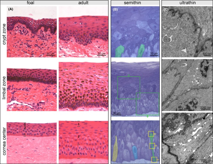
Figure 2.

Representative pictures of crypt zone, limbal zone, and central corneal epithelium illustrating age‐dependent differences (A); in the center of the cornea, a basal (1), intermediate (2), and superficial layer (3) can be distinguished. H&E staining, scale bar =30 µm. Presentation of the total corneal epithelial height (B, center image), detailed view of the intermediate layer (C, upper image) and the basal layer (C, lower image) in a semithin section from the central zone of an adult horse. Toluidine blue staining, scale bar =30 µm and 10 µm respectively. Cells in the intermediate layer have a polygonal shape with horizontal orientation, the suprabasal cell layer being in tight contact with the undulating cell border of the subjacent basal cells (examples marked in green). Three different types of basal cells form a pseudostratified basal layer (highlighted in yellow, blue and green). Cellular details of the cell highlighted in green (yellow boxes) are shown in transmission electron micrographs of ultrathin sections (C) showing the apical interdigitations as well as multiple adhering and occluding junctions at the cell membrane. Scale bar =500 nm and 2000 nm, respectively
