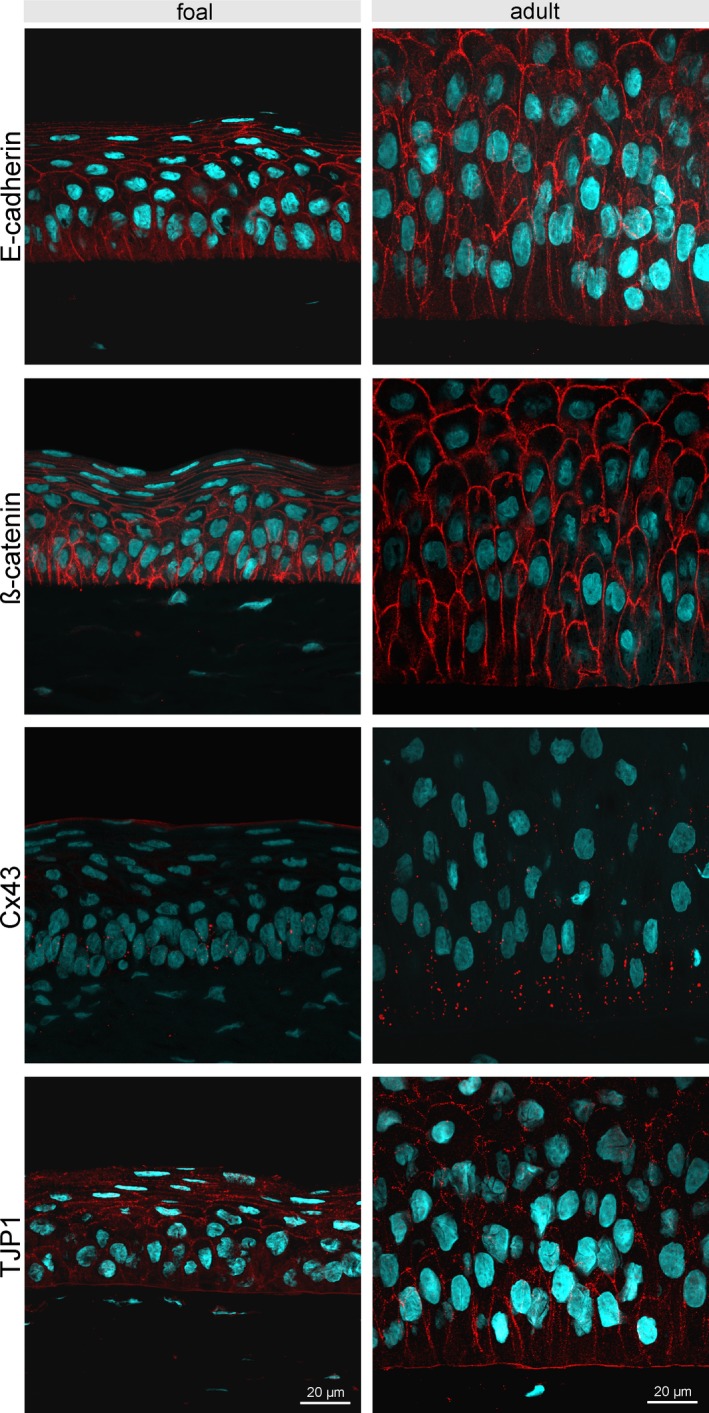
Figure 3.

Immunofluorescent staining of E‐cadherin, β‐catenin, Cx43, and TJP1 of the central corneal epithelium comparing age groups (foal: left panels; adult horse: right panels). E‐cadherin is expressed in the full height of the epithelium, but is not expressed at the area of contact with the basement membrane. Detection of β‐catenin resembles the distribution and localization of E‐cadherin expression. Cx43 is mainly present in the basal epithelial layer. TJP1 was found in all cells including the basal cell membrane. Scale bar =20 µm
