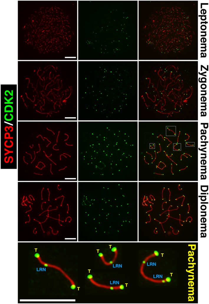
Figure 4.

The pattern of CDK2 localization to chromosomal axes during meiotic prophase I. During leptonema, CDK2 foci (green) can be observed to localize to the telomeres of each chromosome. At this stage, chromosomal axes have not yet been formed as synaptonemal complex formation as determined by SYCP3 staining (red) is still at an early stage. During zygonema, homolog pairing is initiated and distinct axes of SYCP3 can be observed to associate with each other. At this stage, CDK2 localization is still specific to telomeres. During mid‐pachynema, intense singular CDK2 foci can be observed at paired telomeres of homologs. At this point, 1‐2 weaker but distinct interstitial foci of CDK2 can be observed marking the formation of late recombination nodules. In the lower most panel, several examples of paired homologs are shown at higher magnification to indicate both telomeric (T) and late recombination nodule‐associated (LRN) CDK2 foci. At this stage, staining can also be seen on the X‐Y chromosomes, but this is not addressed within the scope of this review. By diplonema, late recombination nodule‐associated CDK2 foci dissociate from the chromosomal axes but can still be observed as pairs of CDK2 foci at chromosomal ends. These represent the individual telomeres of each homolog, which separate upon the splitting of chromosomal axes. Scale bars for each image are shown in white and are equivalent to 5 µm in all pictures.
