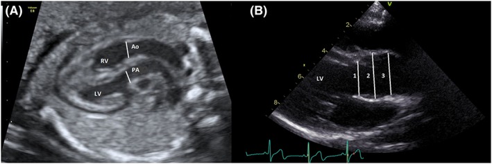
Figure 1.

A, Measurements were made prenatally at the level of the semilunar valve annulus with the valve open. B, Post arterial switch measurements of the aortic root were made in the long axis view with the neo‐aortic valve open; 1 = annulus, 2 = neo‐aortic root, and 3 = sino‐tubular junction. Ao, aorta; LV, left ventricle; PA, pulmonary artery; RV, right ventricle [Colour figure can be viewed at http://wileyonlinelibrary.com]
