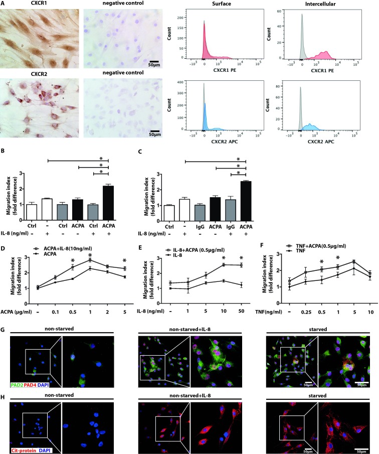
Figure 4.
Collaboration of ACPA-mediated signals with the inflammatory cytokines IL-8 and TNF in inducing FLS migration. CXCR1 and CXCR2 expressions in FLS cultures were analysed with immunohistochemistry by light microscopy with the original magnification of 250x (left panel) and flow cytometry (right panel) (A). Migration of non-starved (B) and starved FLS (C) were assessed after exposing the cells to suboptimal (0.5 µg/mL) dose of ACPA or IgG in the presence or absence of 10 ng/mL recombinant human IL-8. Mean±SD values were calculated from three independent experiments, using cells of five individual patients and three replicates, *p<0.05. Starved FLS mobility was analysed by combining high dose (10 ng/mL) of IL-8 with increasing ACPA concentrations (D) or suboptimal concentration of ACPA with increasing concentrations of IL-8 (E). On similar grounds, increasing concentrations of TNF were combined with ACPA applied at a suboptimal dose (F). Mean±SD values were calculated based on at three independent experiments, using cells of three different patients and six sample replicates, *p<0.05 (G). Confocal microscopy images show increased PAD-4 (red colour) and PAD-2 (green colour) expression of non-starved FLS cultures stimulated by IL-8 (10 ng/mL). Nuclei are represented in blue colour. The effect of IL-8 (10 ng/mL) was also tested on ACPA binding to FLS cultures using confocal microscopy (H). Red colour represents antibody binding, nuclei are shown in blue, The original magnification was 400x. The right panels represent a zoomed area from the original images. ACPA, anticitrullinated protein/peptide antibody; DAPI, 4,6-diamidino-2-phenylindole; FLS, fibroblast-like synoviocytes; IL, interleukin; PAD, protein arginine deiminase; ns, not significant; SF, synovial fluid; TNF, tumour necrosis factor.

