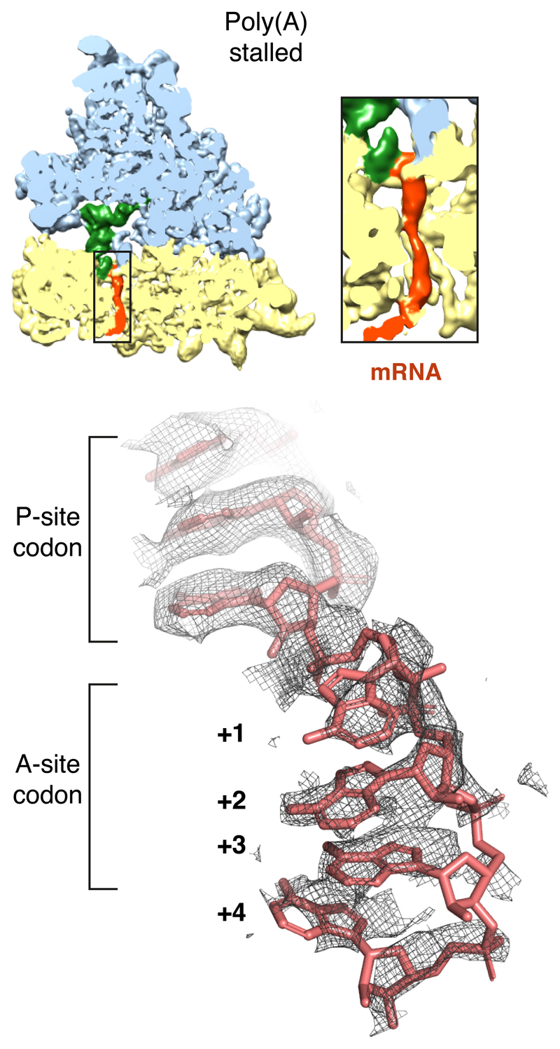
Extended Data Fig. 5. Views of the mRNA density in the EM map of the poly(A)-stalled ribosome.
The density map is sliced through the ribosome in a plane that reveals the decoding centre and shows the mRNA within the small subunit. The large and small subunits (blue and yellow, respectively), P-site tRNA (green) and mRNA (red) are colored. The inset shows a zoomed in region of the mRNA channel, illustrating that the poly(A) mRNA is ordered through most of the channel. The bottom panel shows the mRNA density in the P- and A-sites in the final refined and sharpened map. The mRNA is well ordered in the P-site due to base-pairing with the P-site tRNA, and ordered in the A-site due to stabilizing interactions with rRNA as shown in Fig. 3.

