
Figure 1.
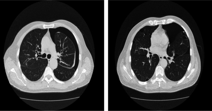
Chest high‐resolution computed tomography of the proband, who experienced recurrent episodes of PSP. Left‐sided PSP and bilateral and multiple bullae are seen as clear. PSP, primary spontaneous pneumothorax
Official websites use .gov
A
.gov website belongs to an official
government organization in the United States.
Secure .gov websites use HTTPS
A lock (
) or https:// means you've safely
connected to the .gov website. Share sensitive
information only on official, secure websites.
Chest high‐resolution computed tomography of the proband, who experienced recurrent episodes of PSP. Left‐sided PSP and bilateral and multiple bullae are seen as clear. PSP, primary spontaneous pneumothorax