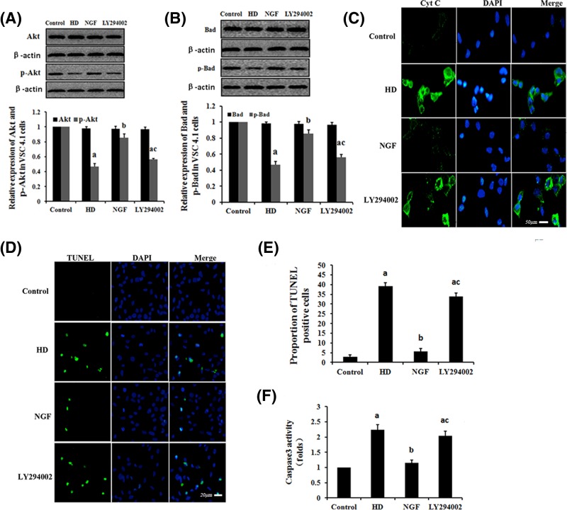
Figure 5. The effect of NGF on HD-induced mitochondrial apoptosis via the PI3K/Akt signaling pathway.
VSC4.1 cells were exposed to 10 mM 2,5-HD, 10 mM 2,5-HD + 50 μg/l NGF or 10 mM 2,5-HD + 50 μg/l NGF + 25 μM LY294002 for 24 h. Western blot analysis was used to detect the Akt, p-Akt, Bad and p-Bad protein expression levels. (A and B) Detection of expression levels of Akt and p-Akt (A) or Bad and p-Bad (B) in VSC4.1 cells, respectively. Data are presented as mean ± S.D. (n=3). (C) Cyt c was examined by immunofluorescence test in each condition (scale bar = 50 μm). (D) TUNEL labeling with DAPI counter staining was performed to detect apoptotic cells in each condition (scale bar = 20 μm). (E) Quantification of the abundance ratio of TUNEL-positive cells in each condition. (F) Detection of caspase-3 activity in each condition. aP<0.05, compared with the control group results; bP<0.05, compared with the results of the group exposed to 10 mM 2,5-HD; cP<0.05, compared with the results of the group exposed to 10 mM 2,5-HD + 50 μg/l NGF.

