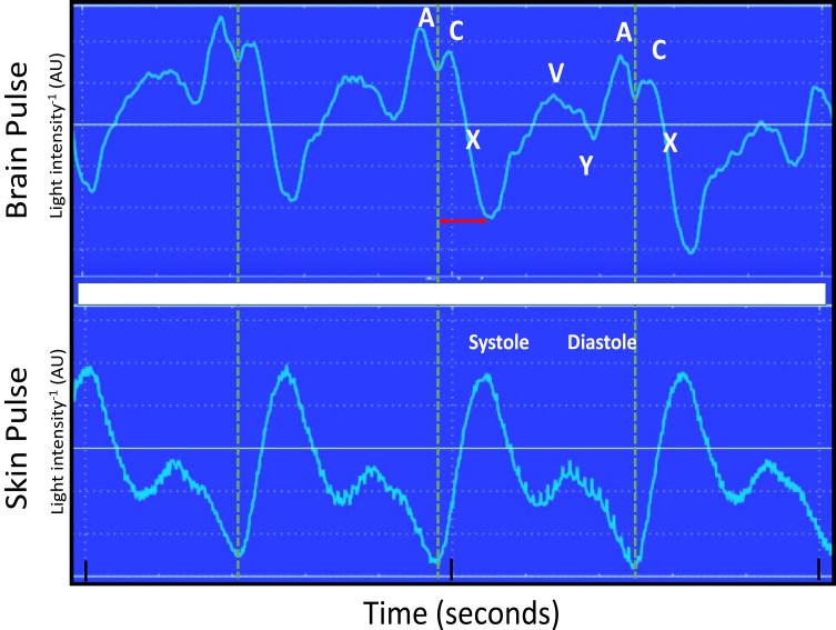
Figure 1.
Simultaneous recording of the brain and conventional skin pulse oximetry waveforms in a single normal sheep brain. The dashed lines represent the start of the skin pulse. The brain and skin pulses were distinct, with the brain pulse demonstrating a waveform similar in shape and timing to a central venous pressure waveform, with A, C, X, V and Y waves, whereas the skin pulse demonstrated a waveform similar in shape and timing to an arterial pressure waveform. In addition, the start of the brain pulse was delayed relative to the skin pulse, by around 100 ms (arrow) and the peak of the pulse was at the end of diastole.
Abbreviation: AU, arbitrary units.

