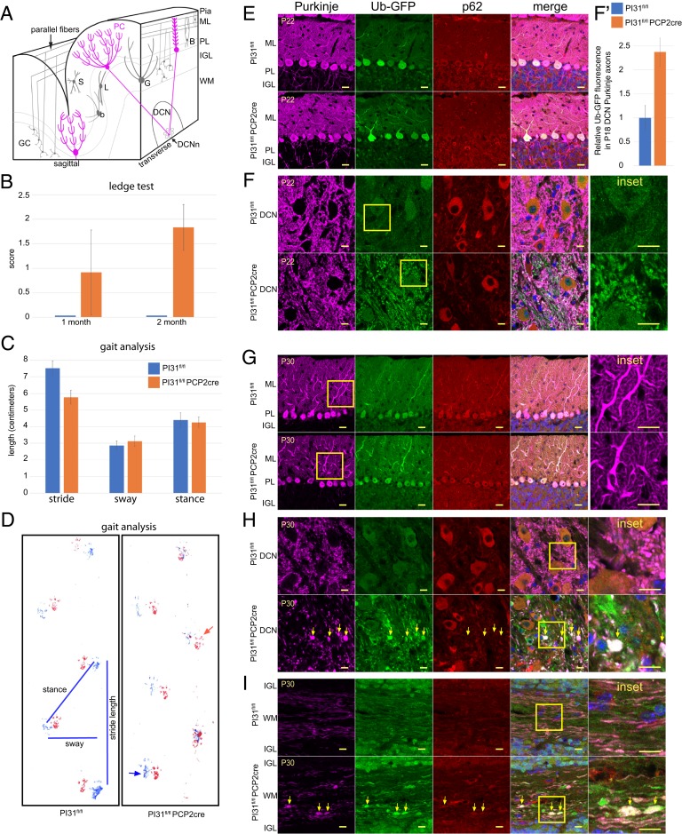
Fig. 4.
Loss of PI31 in PCs leads to aberrant dendrite and axon morphology. (A) Schematic illustrating cerebellar anatomy. PCs (magenta) are the only output of the cerebellar cortex. Their dendrites in the molecular layer (ML) receive inputs from GC parallel fibers and climbing fibers from the inferior olivary nucleus. PC cell bodies form the PC layer (PL), while their axons project through the IGL where mature GCs and Golgi interneurons (G) are found, to the DCN, where they synapse with the DCNn. WM is white matter. (B–D) PI31fl/fl Pcp2-Cre mice display progressive behavioral defects. Up to P22, PI31fl/fl Pcp2-Cre mice were not distinguishable from PI31fl/fl mice in their righting reflex and visual observation of gait. (B) By P30, PI31fl/fl Pcp2-Cre mice began to show loss of balance in the ledge test, which became more severe by P60. The number of mice used for this experiment were: for P30, PI31fl/fl (n = 12) and PI31fl/fl Pcp2-Cre (n = 12), for P60, PI31fl/fl (n = 15) and PI31fl/fl Pcp2-Cre (n = 24). Error bars indicate SD. (C and D) At 10 mo, the gait of PI31fl/fl Pcp2-Cre mice was disturbed, and mutants frequently lost balance. Gait analysis showed a significant decrease in stride length (P = 0.00258 [2-tailed t test), PI31fl/fl [n4] and PI31fl/fl Pcp2-Cre [n4]). Error bars indicate SD. (D) An example of footprints used for gait analysis with measurements of length, sway, and stance. Hind paw prints are blue, and forepaw prints are red. The red arrow points to a common misplaced step for PI31fl/fl Pcp2-Cre mice, while the blue arrow indicates the hind paw touching the surface with splayed toes. (E–I) Cerebellum labeled for Calbindin (magenta) to mark PCs, Ub-GFP (green), p62 (red), and Hoechst 33342 (blue) for nuclei. Yellow box denotes Inset. PI31fl/fl and PI31fl/fl Pcp2-Cre denote PI31fl/fl Ub-GFP and PI31fl/fl Pcp2-Cre Ub-GFP mice, respectively. (E and F) Mature PCs at P22 appeared virtually normal in PI31fl/fl Pcp2-Cre mice when compared to PI31fl/fl mice. (E) PC cell bodies appeared normal, and their dendritic arbors were fully developed. (F) PI31fl/fl Pcp2-Cre mice Purkinje axons in the DCN appeared morphologically normal, but show an increase in Ub-GFP over their control PI31fl/fl siblings. (F′) Level of Ub-GFP increased in P18 PC axons in the DCN of PI31fl/fl Pcp2-Cre mice (significance p – 0.0037 [2-tailed t test], PI31fl/fl [n3] and PI31fl/fl Pcp2-Cre [n3]). Error bars indicate SD (SI Appendix, Fig. S6). (G and H) By P30, PI31fl/fl Pcp2-Cre mice PC primary and secondary dendrites appeared swollen, while their axon terminals in the DCN showed swellings with increased Ub-GFP (yellow arrows), indicating impaired protein breakdown. (I) Loss of PI31 caused swellings (yellow arrows) along the length of the axons in the white matter (P30). (E–H) Single confocal slices. (I) A maximum intensity projection of a confocal stack. (Scale bars: E and G, 20 µm; F, H, and I, 10 µm.)

