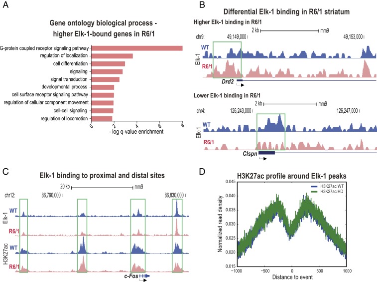
Fig. 3.
Elk-1 binds to genomic sites with H3K27ac signal, and its binding is altered in the 8-wk-old R6/1 striatum. (A) GO analysis of biological processes associated with higher Elk-1 binding in R6/1s compared to wild-type mice. (B) University of California, Santa Cruz (UCSC) Genome Browser images representing normalized Elk-1 ChIP-seq read density from 8-wk-old R6/1 and respective wild-type mice mapped at Drd2 and Claspn example gene loci. Green boxes indicate the Elk-1 occupancy around the TSSs. (C) Tracks from the UCSC Genome Browser displaying normalized Elk-1 and H3K27ac ChIP-seq read density from 8-wk-old R6/1 and respective wild-type mice mapped at a c-Fos example gene locus for proximal and distal regulatory site binding by Elk-1. Green boxes indicate the Elk-1 occupancy around the TSSs and distal sites. (D) H3K27ac profile centering on all Elk-1 peaks shows a valley-shaped chromatin pattern. See also SI Appendix, Fig. S3.

