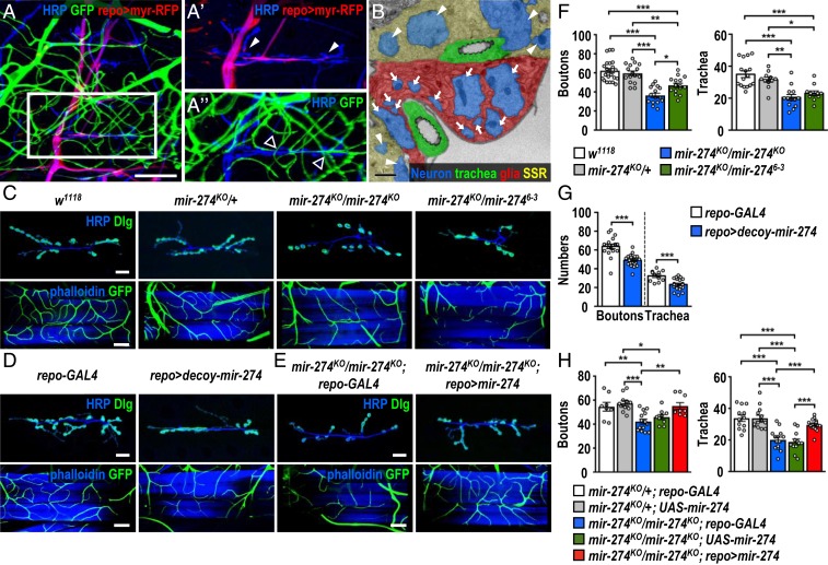
Fig. 1.
Reduced synaptic boutons and tracheal branches in miR-274 mutants. (A to A″) Confocal images show the glia–neuron–trachea system at NMJs of muscle 6/7 for screening miRNA mutants. Larvae were dissected to view glia labeled by repo-GAL4>UAS-myr-RFP, trachea labeled by btl-lexA > lexAop-CD2-GFP, and nerves stained by horseradish peroxidase (HRP). (Scale bar: 30 µm.) Boxed area in A is amplified to show that (A′) glial processes wrap around motor axons but not synaptic boutons (white arrowheads), and (A″) tracheal branches localize close to synaptic boutons (empty arrowheads). (B) Transmission election microscopy micrograph shows that glial processes directly enwrap axons (arrows). Synaptic boutons (arrowheads) are recognized by synaptic vesicles and surrounding SSR. (Scale bar: 1 µm.) (C–E) Images show synaptic boutons (top rows, scale bars: 30 µm) immunostained for presynaptic HRP (blue) and postsynaptic Dlg (green) and tracheal branches (bottom rows, scale bars: 60 µm) revealed by btl-lexA>lexAop-CD2-GFP (green) counterstained for muscle phalloidin (blue), for (C) w1118, mir-274KO/+, mir-274KO/mir-274KO, and mir-274KO/mir-2746−3, (D) repo-GAL4 control (crossed to w1118, Left), and repo-GAL4 crossed to UAS-decoy-mir-274 (repo > decoy-mir274, Right), and (E) the miR-274 mutant carrying GAL4 control (mir-274KO/mir-274KO; repo-GAL4) and glial rescue (mir-274KO/mir-274KO; repo > mir-274). (F–H) Dotted bar graphs for quantification of synaptic boutons and tracheal branches. Data were analyzed by one-way ANOVA followed by Tukey post hoc (F and H) or independent t test (G). See SI Appendix, Table S2. *P < 0.05, **P < 0.01, and ***P < 0.001.

