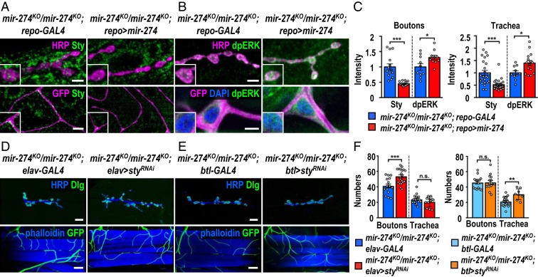
Fig. 6.
Glia-derived exosomal miR-274 modulates synaptic and tracheal growth. (A and B) Confocal images show immunostaining of Sty (A) or dpERK (B) in synaptic boutons (scale bars: 5 µm) and tracheal cells (scale bars: 15 µm in A and 10 µm in B) in mir-274KO/mir-274KO; repo-GAL4 and mir-274KO/mir-274KO; repo > mir-274. Boxed areas are enlarged images. (D and E) Images show specific suppression of synaptic bouton growth in mir-274KO/mir-274KO; elav > styRNAi, compared to mir-274KO/mir-274KO; elav-GAL4 control (D), and specific growth suppression of tracheal branches in mir-274KO/mir-274KO; btl > styRNAi, as compared to mir-274KO/mir-274KO; btl-GAL4 (E). (Scale bars: 30 µm for synaptic boutons and 60 µm for tracheal branches.) (C and F) Dotted bar graphs for quantifications of Sty and dpERK immunofluorescence intensities within synaptic boutons and tracheal cells (C) and numbers of synaptic boutons and tracheal branches (F). See SI Appendix, Table S2. Data were analyzed by independent t tests. n.s., no significance; *P < 0.05, **P < 0.01, and ***P < 0.001.

