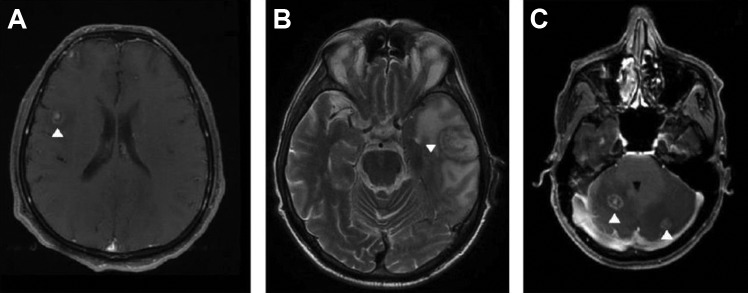
Figure 4.
Magnetic resonance imaging (MRI) of HIV-infected patients with cerebral toxoplasmosis. T1-weighted imaging showed ring-enhancing brain lesion with a small, enhancing asymmetric nodule along the wall of the lesion (the “eccentric target sign; A). T2-weighted imaging showed a lesion with concentric alternating zones of hypo- and hyperintensities (the “concentric target sign”; B). T1-weighted imaging showed ring-enhancing brain lesion with a small, enhancing central nodule (the “target sign”; C). The arrows show the abnormalities.

