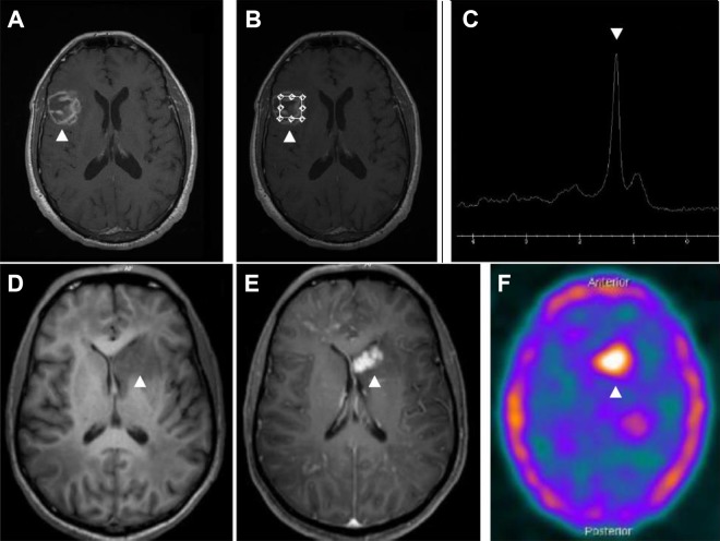
Figure 5.
Magnetic resonance imaging (MRI) and spectroscopy of an HIV-infected patient with cerebral toxoplasmosis (A-C). Gadolinium T1-weighted imaging showing a heterogeneous ring-enhancing brain lesion in right temporoparietal region (B). Spectroscopy image showing increased lipid peak and diminution of other metabolic activity corresponding to the known lesion (C). Magnetic resonance imaging (MRI) and 18F-fluorodeoxyglucose positron emission tomography–computed tomography (18F-FDG PET-CT) image of an HIV-infected patient with histopathologically confirmed primary central nervous system lymphoma (D-F). Gadolinium T1-weighted imaging showed irregular and nodular-enhancing brain lesion in left nucleocapsular region (E). 18F-FDG PET-CT image showing foci of increased metabolic activity corresponding to the known lesion (F). The arrows show the abnormalities.

