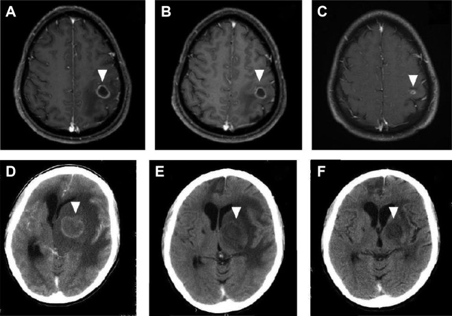
Figure 6.
Magnetic resonance imaging (MRI) of an HIV-infected patient with cerebral toxoplasmosis (A-C). At admission, single lesion was observed in the left parietal lobe (A). After 2 weeks of antitoxoplasma therapy without corticosteroids, partial reduction in both size and perilesional edema was observed (B). After 6 weeks of antitoxoplasma therapy, marked decrease in lesion size and perilesional edema was seen (C). Contrast-enhanced computed tomography (CT) imaging of an HIV-infected patient with cerebral toxoplasmosis (D-F). At admission, extensive single lesion in the left basal ganglia causing brain herniation was seen (D). After 4 weeks of antitoxoplasma therapy with corticosteroids, marked reduction was observed in both size and perilesional edema (E). After 8 weeks of antitoxoplasma therapy, another CT scan showed residual alterations only (F). The arrows show the abnormalities.

