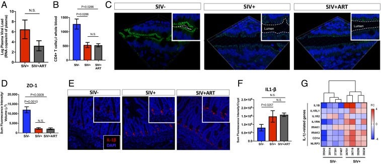
Fig. 1.
Gut epithelial barrier disruption, mucosal CD4+ T cell depletion, and IL-1β expression persist in advanced SIV infection despite late initiation of ART. (A) Viral loads and (B) CD4+ T cell numbers were measured in peripheral blood of SIV-infected rhesus macaques (n = 3) and those treated with ART for >10-wk (n = 5). (C) Immunostaining showed altered distribution of tight junction protein ZO-1 (magnification: 63×) in gut epithelium during SIV infection despite ART, as shown in D semiquantitative analysis by Imaris v8.2. (E) Production of IL-1β (magnification: 20×) from intestinal crypts was increased in SIV infection and did not change after initiation of ART, as shown by (F) semiquantitative analysis on Imaris v8.2. (G) A heatmap dendrogram of IL-1β related genes was analyzed from gene-expression microarray of gut tissue. N.S., not significant.

