Fig. 1.
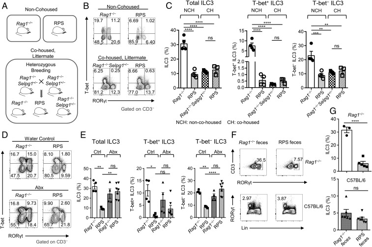
The microbiota from RPS mice induce a transmissible dysbiois and loss of ILC3s. (A) Illustration of mouse housing and breeding scheme. (B) FACS plots of RORγt and T-bet staining gated on CD3‒ LPLs from the large intestine (LI) from non-cohoused (Top) and cohoused (Bottom) RPS, Rag1‒/‒, or Rag1−/−Selpg1+/− mice. (C) Quantification of ILC3 subset frequencies from Rag1‒/‒Selplg+/‒ compared to RPS littermate, cohoused (CH) or non-cohoused mice (NCH). Data are pooled from 2 independent experiments (n = 3 to 4 per group); one-way ANOVA. (D) FACS plots of RORγt and T-bet staining gated on CD3‒ LI LPLs from water-treated (Ctrl; Top) and antibiotics-treated (Abx; Bottom) RPS compared to Rag1‒/‒ mice. (E) Frequencies of ILC3 subsets in antibiotics-treated mice comparing RPS and Rag1‒/‒ mice. Data are pooled from 2 independent experiments (n = 4 to 7 per group); one-way ANOVA. (F) FACS plots of RORγt and CD3‒ staining (Top) or Lineage (Lin) and RORγt staining (Bottom) gated on LI LPLs. (G) Quantification of ILC3 frequencies after indicated feces gavage in the LI of Rag1‒/‒ (Top; n = 3 to 5 per group) and C57BL/6 (Bottom; n = 6 to 7 per group) mice. Data are pooled from 2 and 3 independent experiments, respectively. Error bars indicate SEM. *P < 0.05, **P < 0.01, ***P < 0.001, ****P < 0.0001; ns, not significant.

