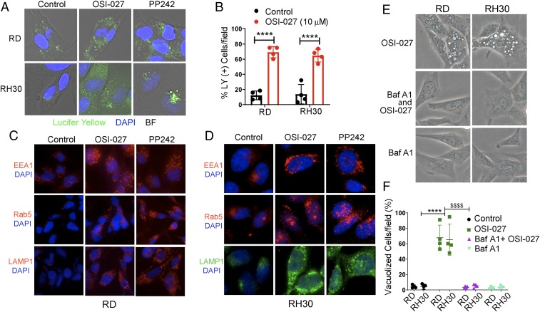
Fig. 3.
Vacuoles induced by dual mTORC1/2 inhibitors are macropinosomes. (A) Overlay of bright field microphotographs on LY accumulation in untreated and OSI-027– or PP242-treated RD or RH30 cells at 24 h (also see SI Appendix, Fig. S3A). The majority of the vacuoles were positive for LY. The nature of the unstained vacuoles was not characterized. (B) Quantification of OSI-027–treated (10 µM, 24 h) LY-positive RD and RH30 cells compared with vehicle-treated control cells. (C and D) Fluorescence immunostaining for EEA1, Rab5, and LAMP-1 in RD (C) and RH30 (D) cells treated with vehicle (control), OSI-027 (10 µM, 24 h), or PP242 (2.5 µM, 24 h) (also see SI Appendix, Fig. S3 C and D). (E and F) Phase-contrast images (E) and bar graph (F) showing that BafA1 pretreatment (0.1 µM, 1 h) of RD and RH30 cells almost completely abrogated OSI-027–induced vacuolization. (Magnification: A and D, 400×; C and E, 200×.) ****P < 0.0001 compared with controls; $$$$P < 0.0001 compared with OSI-027. Four to 5 fields of 50 to 100 cells/field for each treatment were counted. This experiment was repeated twice.

