Fig. 7.
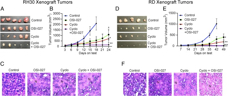
OSI-027 enhances the inhibitory efficacy of cyclophosphamide against RMS cell-derived xenograft tumors. Tumors were derived from RH30 cells (A–C) or RD cells (D–F). Tumor volumes were ∼80 mm3 when treatments started. (A and D) Representative tumors excised from vehicle- or drug-treated animals (n = 5). (B and E) Line graphs showing the inhibitory effects of OSI-027 (75 mg/kg orally, 3 times/wk), cyclophosphamide (Cyclo) (60 mg/kg, i.p., 2 times/wk), or both on the growth of xenograft tumors. (C and F) Hematoxylin and eosin staining of the 5-µm sections of formalin-fixed xenograft tumors. (Magnification: A, 100×; B and C, 200×.) Tumor sections from vehicle-treated control animals show highly condensed areas of proliferating cells with dark nuclei, whereas the sections from OSI-027–treated and/or cyclophosphamide-treated animals show disrupted tumor tissue architecture associated with necrotic patches (N). *P < 0.05; **P < 0.01; ***P < 0.001 compared with vehicle-treated control tumors. Each set of data represents n = 5.

