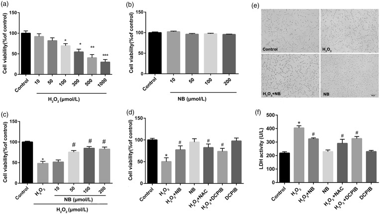
Figure 1.
Effect of NB on H2O2-induced neuronal injury. (a) HT22 neuron cells were incubated with different concentrations H2O2 for 24 h, and cell viability was measured using CCK8 kit; (b) HT22 neuron cells were treated with different concentrations NB for 24 h and the cell viability was measured; (c) Cells were pretreated with different concentrations of NB or vehicle alone for 2 h and were then treated with 300 µmol/L H2O2 for 24 h before cell viability detection. (d) Cells were pretreated with 100 µmol/L NB or 10 µmol/L DCPIB for 2 h followed by the further incubation of 300 µmol/L H2O2 for 24 h. N-acetyl-L-cysteine (NAC) at 100 µmol/L was used as a positive control for indicating the anti-oxidative activity. (e) Morphology of cells in response to treatment of NB and H2O2. Bar = 50 µm. (f) LDH release in the supernatant of HT22 cells treated with NB or DCPIB followed by H2O2. *P < 0.05, **P < 0.01, ***P < 0.001 vs. the control group; #P < 0.05 vs. the H2O2 group.

