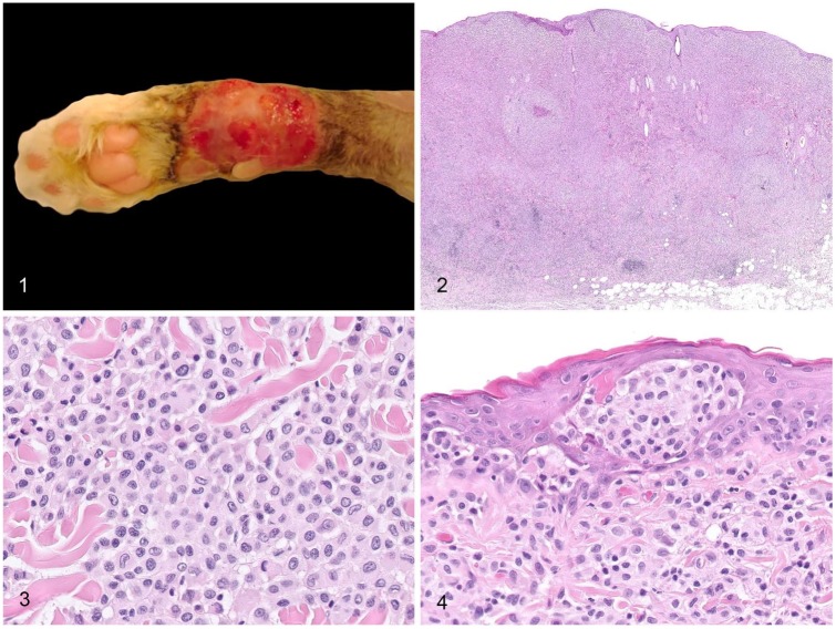
Figures 1–4.
Gross and histologic features of feline progressive histiocytosis skin nodules. Figure 1. Large alopecic and ulcerated plaque on the distal limb of a cat. Image provided by MicenVet. Figure 2. Cutaneous and subcutaneous densely cellular, unencapsulated, and poorly circumscribed histiocytic nodule, with clusters of reactive lymphocytes at the deep periphery of the lesion. H&E. Figure 3. The atypical cells are round-to-polygonal with distinct cell borders, abundant pale eosinophilic homogeneous to finely granular cytoplasm, and a central-to-paracentral, oval-or-indented nucleus with marginated chromatin. Anisocytosis and anisokaryosis are mild-to-moderate. H&E. Figure 4. Skin nodule with intraepidermal aggregates of atypical cells (epitheliotropism). H&E.

