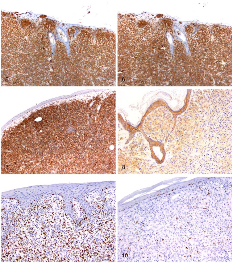
Figures 5–10.
Immunohistochemical features of feline progressive histiocytosis skin nodules. Immunohistochemistry; chromogen: 3,3’-diaminobenzidine, hematoxylin counterstain. Figure 5. Dermal atypical cells are diffusely positive for MHC II. Figure 6. Dermal atypical cells are diffusely positive for Iba1. Figure 7. Dermal atypical cells are diffusely positive for CD18. Figure 8. Dermal and epitheliotropic atypical cells are positive for E-cadherin. Figure 9. Dermal atypical cells have a Ki67 index > 15% (~60%). Figure 10. Dermal atypical cells have a Ki67 index ≤ 15% (~5%).

