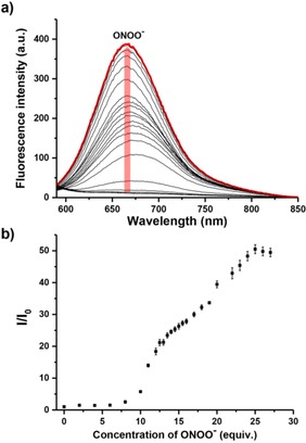
Figure 1.

(a) Fluorescence spectra of DCM‐Bpin (10 μM) with addition of ONOO− (from 0 to 27 equiv.) in PBS buffer solution (10 mM, containing 5 % DMSO, pH=7.40). The red line shows the highest intensity after addition of ONOO− (25 equiv.); (b) Fluorescence intensity changes (I/I0) of probe DCM‐Bpin (10 μM) with addition of ONOO− (from 0 to 27 equiv.) in PBS buffer solution (10 mM containing 5 % DMSO, pH=7.40) after 5 min. λex=560 nm/λem=667 nm. Slit widths: ex=10 nm, em=20 nm.
