Figure 1.
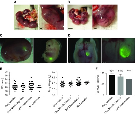
Transplantation of renal progenitor cells into the fetus allows surviving without inhibiting growth. (A) Photomicrographs of a fetus in utero at E13.5. (B) Fetus in utero after cutting the uterine muscle layer. (C and D) Visualization of GFP-expressing transplanted RPCs in the retroperitoneal cavity at E13.5. Images demonstrate a fetus removed after cell injection and dissected for validation of cell transplantation. (E, left) Crown-rump length (CRL) and (right) body weight at E19.5 were compared across the RPC-injected experimental group (n=35), a noninjected (puncture only) group (n=15), and a sham-operated group (n=20). (F) The survival rate was calculated as a percentage of the number of surviving fetuses 6 days after the operation (sham operation, n=14; puncture only, n=20; and RPC injection, n=19). There was no significant difference between groups. Bars represent means±SEM. P<0.05, Mann–Whitney U test. Scale bars, 1 mm in (A), (B), and left panel of (C) and (D); 2 mm in right panel of (C); 250 µm in right panel of (D).

