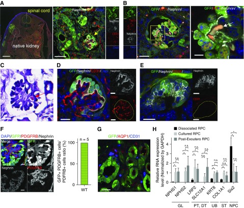
Figure 2.
Analysis of nephrons derived from RPC regenerated inside the retroperitoneal cavity of fetus. (A) In immunofluorescence imaging, transplanted RPCs expressing GFP were present outside the host kidney in the retroperitoneum. Mature glomeruli with capillary loops (yellow arrow) derived from transplanted cells can be seen. (B and C) Erythrocytes (white arrow in [B] and red arrow in [C]) and blood flow were identified. (C) Hematoxylin and eosin stain. (D) In vivo glomeruli had capillary loops containing host embryo vascular endothelial cells (CD31+ and GFP–). (E) In vitro glomeruli without vascular endothelial cells (CD31+). (F) PDGFRb-positive cells in GFP-expressing glomeruli. All PDGFRb-positive cells expressed GFP. Refer to Supplemental Methods. (G) Transplanted cells expressing tubule markers AQP1 (red). (H) Quantified PCR analysis of nephron genes. Kruskal–Wallis test was for comparison. Error bars indicate SEM; *P<0.05; n=6. Scale bars, 500 µm in the left panel of (A); 50 µm in the right panel of (A); 20 µm in left panel of (B) and (C); 5 µm in right panel of (B); 20 µm in (D), (E), and (G); 10 µm in (F). DAPI, 4′,6-diamidino-2-phenylindole; DT, distal tubule; GAPDH, glyceraldehyde-3-phosphate dehydrogenase; GL, glomerulus; PT, proximal tubule; ST, stromal tissue; WT, wild type.

