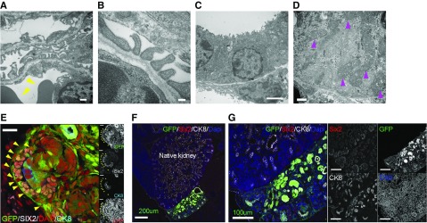
Figure 3.
Nephrons regenerate inside the retroperitoneal cavity are highly differentiated but do not integrate with the host kidney beyond the renal capsule. (A and B) Podocytes and slit diaphragms in glomeruli derived from transplanted cells. Erythrocytes indicated by yellow triangles on the endothelial side. (C) Brush border in the lumen of the proximal tubule. (D) There were abundant mitochondria in the cells (magenta triangles). (E) GFP-positive cells surrounded Six2-positive NPCs and Six2-negative cells (yellow triangle) in the cap mesenchyme. (F and G) Donor UB cells differentiated into UB tip and collecting duct but did not integrate with the host UB. Scale bars, 500 nm in (A) and (D); 100 nm in (B); 2 µm in (C); 20 µm in (E); 200 µm in (F); 100 µm in (G). CK8, Cytokeratin 8; DAPI, 4′,6-diamidino-2-phenylindole.

