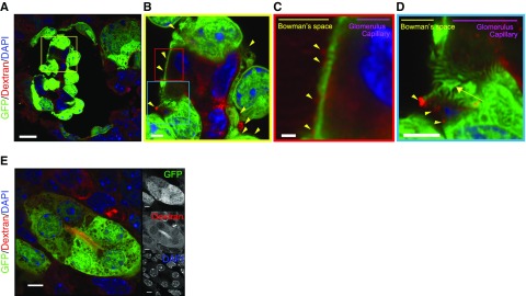
Figure 4.
Generated glomeruli displayed podocytes and filtration function. (A–C) Fluorescence-labeled dextrans in blood were filtered to the Bowman’s space side in the glomerulus (displayed filtered dextran with yellow triangles) derived from RPCs implanted in the wild-type mice. (D) Podocytes formed a filtration slit (yellow arrow). (E) Dextran was seen in the uriniferous tubule cavity. Scale bars, 20 µm in (A); 2 µm in (B) and (D); 1 µm in (C); 5 µm in (E). DAPI, 4′,6-diamidino-2-phenylindole.

