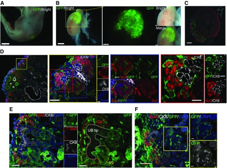
Figure 5.
Transplantation of RPC under the renal capsule enables integration with the host kidney but is a chimeric structure. (A) Fetus at E19.5 with RPCs expressing GFP (day 6 postinjection). (B) GFP-expressing progenitor cells integrated into the host kidney. (C and D) Integration of exogenous NPCs into host cap mesenchyme. Higher magnification of (D) left panel (blue frame panel) showing transplanted NPCs in the host renal vesicle (RV), and (red frame panel) chimerism of NPCs in cap mesenchyme (CM) visualized by GFP. (E) Mosaicism in the RV. (F) Chimeric glomeruli-like mosaics contributed by the host and donor NPCs. The yellow dotted line indicates the glomerulus. Scale bars, 2 mm in (A); 1 mm in left panel of (B); 250 µm in right panel of (B); 200 µm in (C); 20 µm in left panel and yellow-framed panel of (D) and in (E); 10 µm in blue- and red-framed panel of (D); 50 µm in (F). CK8, Cytokeratin 8; DAPI, 4′,6-diamidino-2-phenylindole.

