Figure 7.
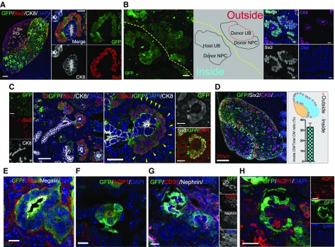
RPC transplanted in a renal-deficient fetus connect with the host ureteric bud to form nephrons. (A) Immunolabeling of transplanted cells integrated into the host kidney. Transplanted NPCs gathered around the UB tip and formed cap mesenchyme (CM). (B) Transplanted cells present outside the kidney self-organized to form CM with GFP-expressing UB and NPCs (outside). The transplanted cells that reached the host nephrogenic zone aggregated around the host UB tip (GFP negative) and formed new CM (inside). (C) GFP-negative cells (yellow triangle) surrounded Six2-positive NPCs in the CM. (D) Overall, 33% of the regenerated cap mesenchyme was integrated with the host UB tip (counted to a total of 119 tips, n=4 slices). Refer to Supplemental Methods. (E–H) Transplanted cells expressing tubule and nephron markers. (G and H) Mature glomeruli with capillary loops derived from transplanted GFP cells. (H) AQP1 is expressed in structures associated with the developing renal vasculature. The expression of AQP1 was observed inside regenerated glomeruli. It was noted as a host-derived tissue because it was GFP negative. Scale bars, 100 µm in (A) and (B); 50 µm in (C); 10 µm in (E) and (F); 20 µm in (G) and (H). CK8, Cytokeratin 8; DAPI, 4′,6-diamidino-2-phenylindole.

