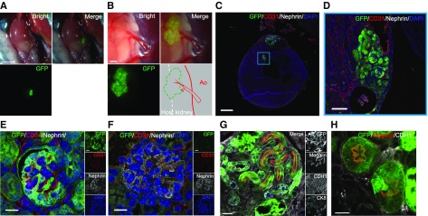
Figure 8.
Regenerating nephrons inside the retroperitoneal cavity of the Wild-type mouse are able to be maintained after birth. (A and B) Transplanted GFP-RPCs adhered to the native kidney and were connected to the host’s blood vessels. (C) Kidney was collected and immunofluorescence staining was performed. (D) Transplanted GFP-RPCs partially penetrated into the fetal kidney at postnatal day 14 (P14). (E) The transplanted GFP-RPCs differentiated into nephrons with expressed nephrin, and CD31-positive endothelial cells were observed in the glomerulus under high magnification (63× magnification). (F) As control, a normal B6 fetal glomerulus on P14. (G) The transplanted GFP-RPC expressed a proximal tubular marker (Megalin). A site positive for CDH1 (collecting duct and distal tubule marker) and negative for CK8 (collecting duct marker) indicates a distal tubule. (H) Proximal tubule in the high magnification. Scale bars, 2 mm in (A); 250 µm in (B); 500 µm in (C); 20 µm in (D) and (G); 10 µm in (E), (F), and (H). Ao, Aorta; CDH1, Cadherin 1; CK8, Cytokeratin 8; DAPI, 4′,6-diamidino-2-phenylindole.

