Figure 6.
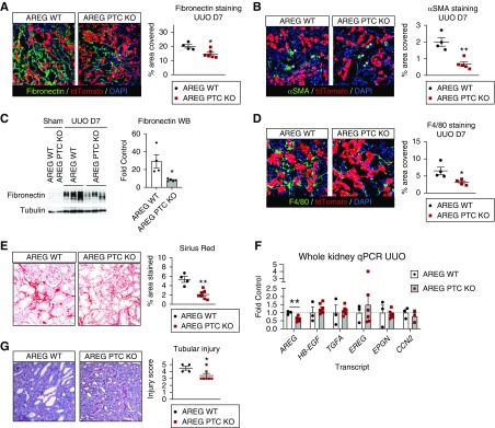
AREG PTC-KO reduces kidney fibrosis after UUO. AREG PTC-KO mice or AREG WT littermates were subjected to UUO and kidneys were collected after 7 days. (A and B) The profibrotic markers fibronectin and αSMA were examined by immunofluorescence staining (left panels) and the levels of the stained area per field for each marker (% area covered, right panel graphs) were quantified. (C) The levels of fibronectin are examined in whole kidney lysates by Western blot (left panels, tubulin is used as loading control) and quantified by densitometric analysis (right panel graph). (D) Macrophage infiltration was examined by F4/80 staining (left panels) and the levels of the stained area per field (% area covered, right panel graphs). (E) Collagen deposition was quantified after Sirius red staining. (F) Whole kidney mRNA was extracted and the expression of AREG, HB-EGF, TGFA, epiregulin, epigen, and CCN2 was tested by quantitative PCR and presented after normalization to AREG WT mice subjected to UUO (fold control). (G) Tubular injury was scored in kidney sections after periodic acid–Schiff staining as described under Methods. Representative images ([G], left panel) and scoring results ([G], right panel) are shown. n=4–7; *P<0.05; **P<0.01.

