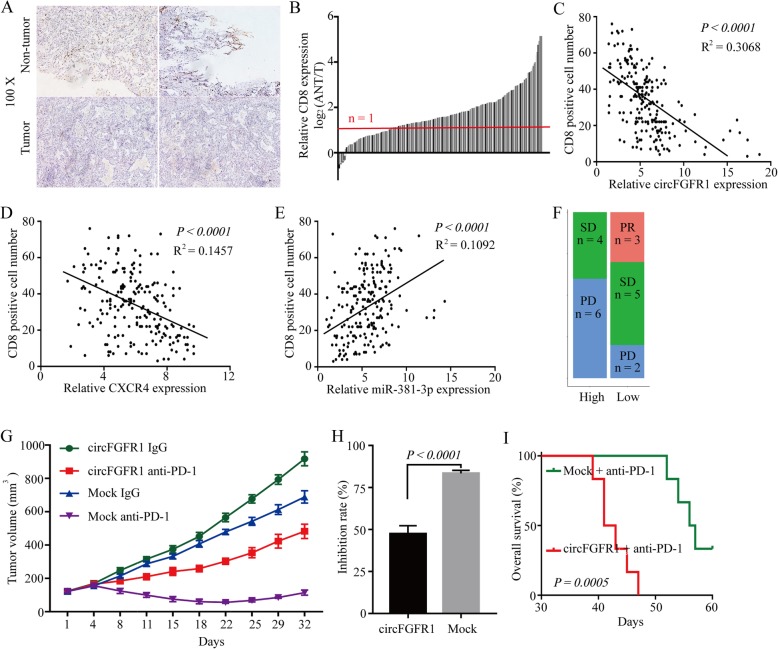
Fig. 6.
CircFGFR1 promotes immunosuppression and resistance to NSCLC immunotherapy. a Representative NSCLC cases in which the tissue were analyzed by IHC staining for CD8. b CD8+ T cells in 210 pairs of NSCLC and matched nontumor tissues, shown as log2 (tumor/nontumor). c A negative correlation between circFGFR1 and the number of CD8-positive cells was observed in the NSCLC tissues (R2 = 0.3068; P < 0.0001). d A negative correlation between CXCR4 and the number of CD8-positive cells was observed in the NSCLC tissues (R2 = 0.1457; P < 0.0001). e A positive correlation between miR-381-3p and the number of CD8-positive cells was observed in the NSCLC tissues (R2 = 0.1092; P < 0.0001). f Comparison of therapy efficacy for patients with high and low circFGFR1 expression and who were treated with Opdivo. g LLC-mock or LLC-circFGFR1 cells were subcutaneously injected into 4-week-old nude mice, and when tumors reached a mean tumor volume of 100 mm3, the mice were treated with an IgG or PD-1 antibody. The data are expressed as the mean tumor volume. h The data are expressed as the percent of tumors with inhibited growth (the data are presented as the mean ± SD). i Comparison of the overall survival curves for mice with high and low circFGFR1 expression of xenograft lung tumors that were treated with a mouse antibody against mouse PD-1

