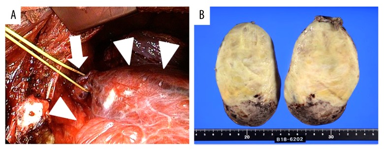
Figure 3.
(A) Intraoperative findings revealed a gigantic mass (arrow heads) adhering to the chest wall via a pedicle (arrow). The mass and its pedicle were completely resected thoracoscopically. (B) The macroscopic appearance was an oval, yellowish-white tumor with a diameter of 10 cm or more, and the tumor had hemorrhage and necrosis on the caudal side, consistent with the lesion showing inhomogeneous intensities in the chest CT.

