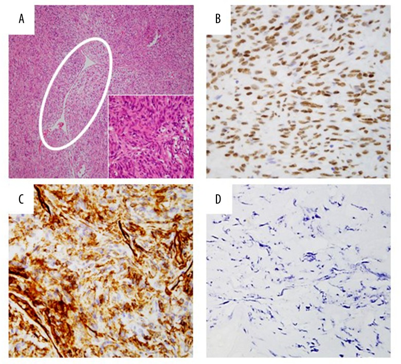
Figure 4.
Histological specimen revealed irregular fascicles of spindled cells and staghorn-like blood vessel (white circle) with deposition of collagen (A). Immunohistochemical staining of tumor cells were positive for CD34 (B) and STAT6 (C). Spindle cells proliferation and STAT6-positive cells were not observed in the pedicle. Ki-67 (MIB-1) proliferation index was less than 1% (D).

