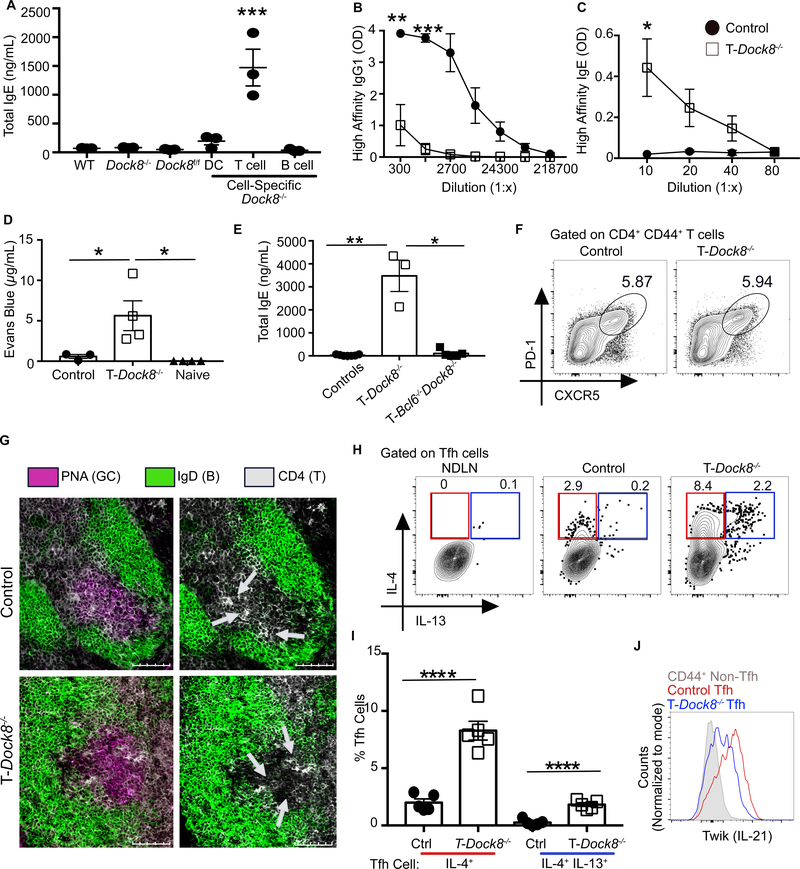
Fig. 1. DOCK8 deficiency reveals the presence of a specific Tfh cell population associated with a hyper-IgE state.
(A) WT, Dock8−/−, Dock8fl/fl, Cd11cCreDock8fl/fl (DC-Dock8−/−), Cd4CreDock8fl/fl (T-Dock8−/−), and Cd19CreDock8fl/fl (B-Dock8−/−) mice were immunized intranasally (i.n.) with LPS and NP16-OVA (LPS+OVA). Day 12 total serum IgE was measured by ELISA. (B and C) T-Dock8−/− or control mice (Dock8fl/fl) were immunized and boosted with LPS+OVA. Day 8 post-boost sera were analyzed by ELISA for (B) NP4-specific IgG1 and (C) NP4-specific IgE. OD, optical density. (D) PCA assay performed by transferring day 8 post-boost sera into naïve recipients and challenging with NP7-BSA and 1% Evans blue. Dye extravasation quantification is shown. (E) Day 12 serum IgE from T-Dock8−/−, T-Bcl6−/−Dock8−/−, or control mice (Dock8fl/fl and Bcl6fl/fl Dock8fl/fl) immunized i.n. with LPS+OVA. (F) Day 8 Tfh cell frequencies are depicted as representative flow cytometry contour plots. (G) Immunofluorescent images of day 9 MedLN GCs from immunized T-Dock8−/− or control Dock8fl/fl mice stained for IgD (green) and CD4 (white) with or without (left and right, respectively) peanut agglutinin (PNA, purple) are shown. Scale bars: 100 μM. Arrows indicate clusters of CD4+ T cells in the GC. (H and I) Intracellular expression of IL-4 and IL-13 by day 8 Tfh cells (gated as in fig. S4A) depicted as (H) flow cytometry plots and (I) bar graphs. NDLN, nondraining lymph node. (J) IL-21 reporter expression in Tfh cells from IL-21 TWIK TDock8−/− or control Dock8fl/fl reporter mice on day 8 after-immunization depicted as histogram overlay. In (A), (D), (E), and (I), each symbol indicates an individual mouse. Numbers in flow plots indicate percentages. Error bars indicate SEM. Statistical tests: analysis of variance (ANOVA) (A and D); Student’s t test (B, C and I); Kruskal–Wallis H test (E). *P<0.05, **P<0.01, ***P<0.001, ****P<0.0001. Data representative of at least two independent experiments with three to seven mice per group.

