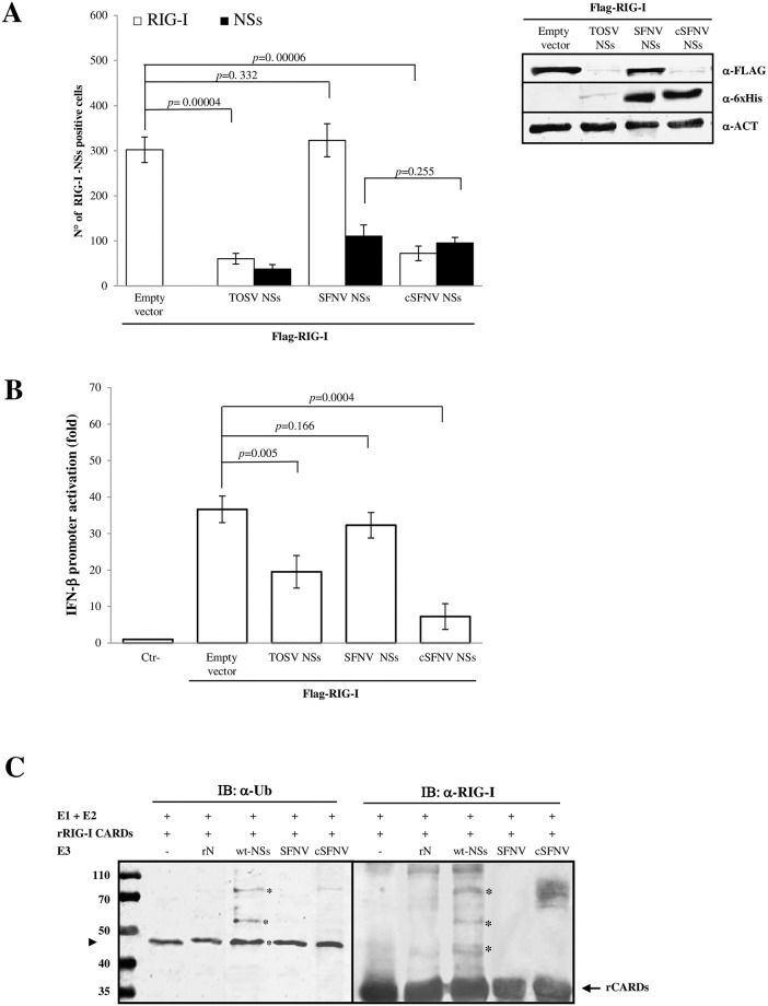
Fig 2. C-terminus of TOSV NSs is linked to E3 ubiquitin ligase activity.
The fusion of TOSV NSs C-terminus to Sandfly Fever Naples Virus (SFNV) NSs conferred it a different behaviour. (A, left panel) The chimeric protein cSFNV NSs was tested for its degrading activity on RIG-I co-transfected cells, by immunofluorescence, using specific antibodies [RIG-I (□), NSs (■)]. Graphs are based on the mean values of three independent experiments ± SD. (A, right panel) A more accurate analysis of cellular RIG-I degradation was performed in co-transfected cells by immunoblotting on the whole cell lysates using anti-FLAG or anti-6xHis antibodies. The intensity of the RIG-I band was quantified by densitometry (Supplement data 3). (B) Lenti-X 293T cells were transfected with IFN-β reporter plasmid, FLAG-RIG-I expression plasmid along with TOSV wt-NSs, SFNV wt-NSs or chimeric cSFNV NSs expressing plasmids. Luciferase activities were measured after poly(I:C) treatment. Fold induction was calculated for each sample with respect to the basal empty plasmid transfected sample, after normalization of the signal with the pSV40-RenLuc internal control. The mean values of at least three sets of experiments ± SD are presented. C) cSFNV NSs showed E3 ubiquitin ligase activity in the biochemical reaction, in association with UbcH5b/c as E2. Poly-ubiquitinated bands of rRIG-I CARDs were detected by immunoblotting using anti-RIG-I or anti-Ub antibodies when cSFNV or TOSV NSs was used in the reaction in place of E3 Ub ligase. No ubiquitination activity was shown when SFNV NSs was used.

