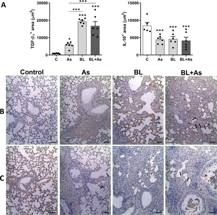
Fig 5. Immunohistochemical characterization of TGF-β1 and IL-10 expression in the lung parenchyma related to Ascaris suum infection, pulmonary fibrosis and comorbidity in mice.
(A) morphometry of TGF-β1 expression in the lungs and morphometry of the positive area of IL-10 expression in the lungs. (B) Representative immunohistochemestry for TGF-β1 expression in the lungs. (C) Representative Immunohistochemistry for IL-10 in the lungs, Results represent mean ± S.E.M., *P< 0.05, **P< 0.01, ***P< 0.001, or One-way ANOVA test was used. Ascaris suum larvae depicted by arrows, Bar = 100μm.

