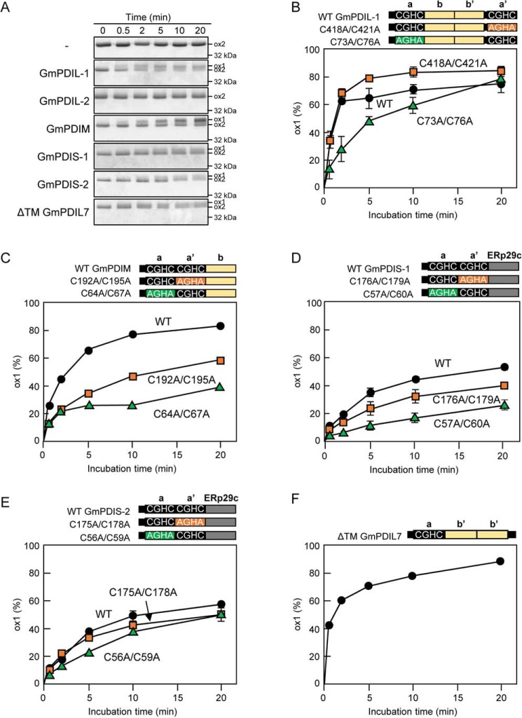
Figure 5.
Conversion of ox-2 GmERO1a to the ox-1 form by reduced PDI family proteins. A, GmERO1a (5 μm) was incubated with 2 μm reduced GmPDIL-1, GmPDIL-2, GmPDIM, GmPDIS-1, GmPDIS-2, or ΔTM GmPDIL7 in the presence of 10 mm GSH at 25 °C, then treated with N-ethylmaleimide, and subjected to nonreducing SDS-PAGE. Proteins were stained with Coomassie Brilliant Blue G-250. B–F, schematic representations of WT PDI family proteins and respective mutants are shown. Numbers in the designations of mutants indicate cysteine residues substituted with alanine residues. GmERO1a and each PDI family protein (circles) or respective domain a (green triangles) or domain a′ (orange squares) active-center cysteine mutant was incubated and separated as described in A. The percentage of GmERO1a in the ox-1 form was calculated based on the ox-1 and ox-2 band intensities. Data are presented as the mean ± S.E. of n = 3 experiments.

