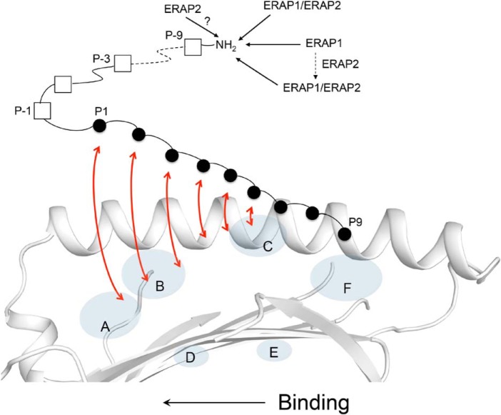
Figure 7.
Trimming of MHC I–bound 18-mer precursor peptide by the ERAP enzymes. Shown is a model of antigen processing in which an N-terminally extended candidate peptide is bound into the MHC I groove by only a few C-terminal residues. As the peptide undergoes a dynamic binding and “sampling” into the groove (indicated by red arrows), from its C to N terminus, the N-terminal residue extensions are concurrently trimmed by the ERAPs. Inside the cells, the ERAP1 and ERAP2 enzymes likely exist in more than one molecular form, with each form shaping differently the MHC I immunopeptidome (see “Discussion”). ERAP1/ERAP2 indicates the heterodimer.

