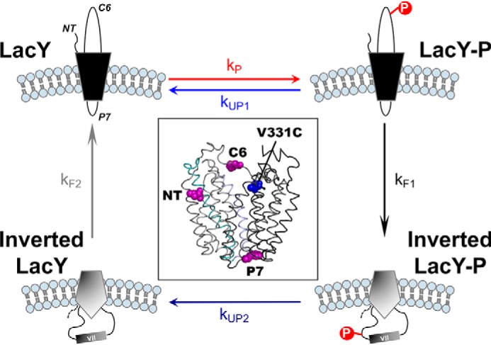
Figure 1.

Schematic of the events occurring during phosphorylation/dephosphorylation-induced topological switching of a membrane protein in proteoliposomes. LacY topology is shown when assembled in proteoliposomes containing E. coli native lipids (correct LacY, top left) before and during phosphorylation by a kinase located outside of the proteoliposomes. The NT, C6, and P7 domains are indicated. Membrane phospholipids are depicted in light blue. LacY-P, phosphorylated LacY. The boxed area depicts a side view of LacY (PDB code 2CFQ) with the diagnostic Trp replacements introduced in EMD NT, C6, and P7 (magenta spheres) and the IAEDANS label at Cys-331 (blue spheres).
