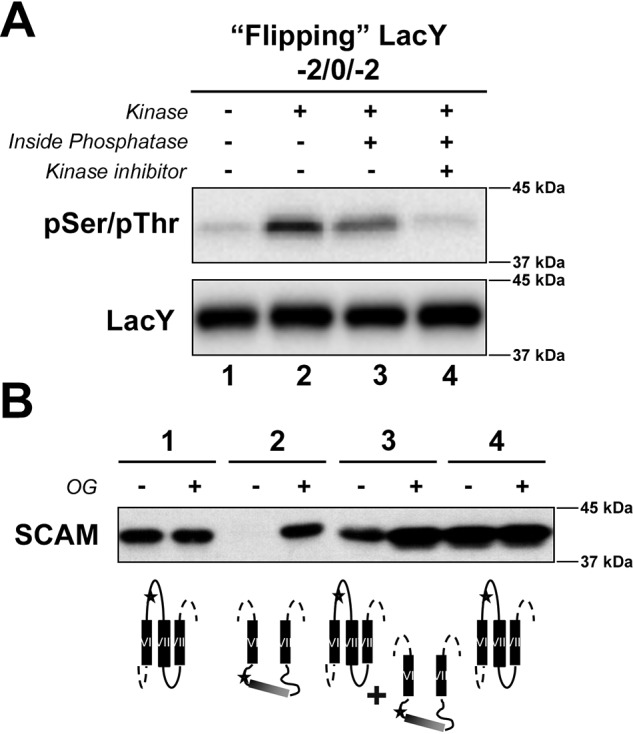
Figure 2.

Determination of the reversibility of phosphorylation-induced topological switch of a membrane protein upon dephosphorylation in the proteoliposomes at steady state. A, determination of the phosphorylation state of the LacY template −2/0/−2 containing a single engineered PDK1 sequence in EMD C6 in the presence or absence of external kinase and its inhibitor, with and without encapsulated phosphatase, as indicated. LacY was reconstituted in proteoliposomes made of E. coli native lipids. The phosphorylation state of LacY was visualized by Western blotting using an anti-pSer/pThr antibody after quenching in Laemmli buffer. B, determination of the orientation of EMD C6 of LacY with altered EMD net charge and containing a single cysteine replacement using SCAMTMD under the various conditions indicated in A. Proteoliposomes were exposed to MBP before or after addition of OG. The deduced orientation of EMD C6 is also shown. The orientation of LacY TMDs VI–VIII is summarized for LacY in proteoliposomes. Stars indicate the position of single Cys replacements in EMD C6 (H205C), used to determine topological orientation.
