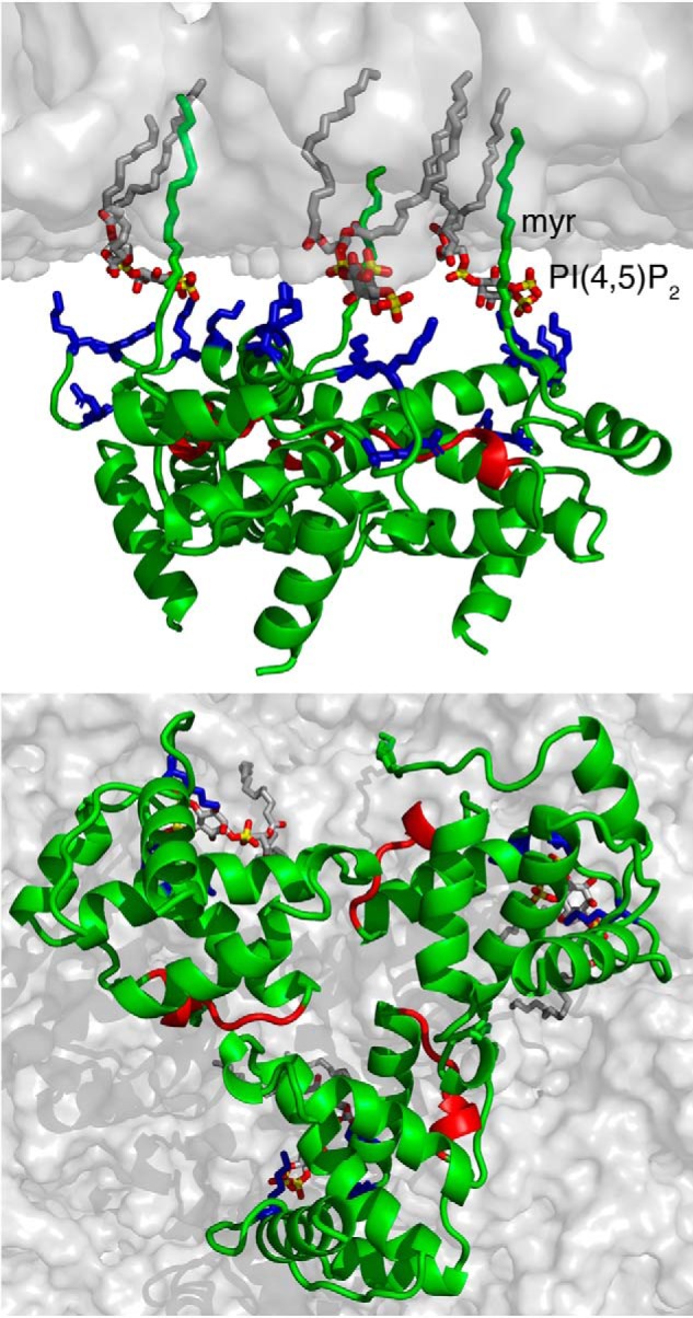
Figure 10.

A model of HIV-1 MA trimer bound to membrane. Shown are top and bottom views of the MA trimer bound to a membrane bilayer. The interaction is mediated by the myr group, the acidic polar head of PI(4,5)P2, and basic residues (Arg22, Lys26, Lys27, Lys30, and Lys32) in the HBR (blue). Residues colored in red are located in the trimeric interface, as revealed by the HDX-MS data. Membrane bilayer was generated in the VMD membrane builder plug-in (92). PI(4,5)P2 was generated in Avogadro (93). The MA trimer was constructed by superimposition of the structure of myr-exposed MA and the X-ray structure of myr(−)MA.
