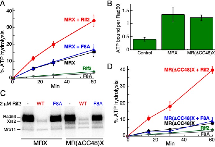
Figure 4.

Rif2 stimulates the ATPase activity of MRX. A, standard ATPase assays with DNA and WT MRX contained either no Rif2 (black), 2 μm Rif2 (red), or Rif2-F8A (blue). Negative controls contained 2 μm Rif2 (green) or Rif2-F8A (gray), but no MRX. B, nitrocellulose filter binding assays of [γ-32P]ATP to WT or mutant MRX, as described (36). Control, no MRX. Quantification of three independent experiments with S.E. is shown. C, Tel1 kinase assays with 30 nm MRX or M(Rad50ΔCC-48)X and 2 mm Rif2 or Rif2-F8A as indicated. D, ATPase reactions as in A, but with M(Rad50ΔCC-48)X. The green Rif2 and gray Rif2-F8A curves, minus MRX, are from the same data set as shown in A.
