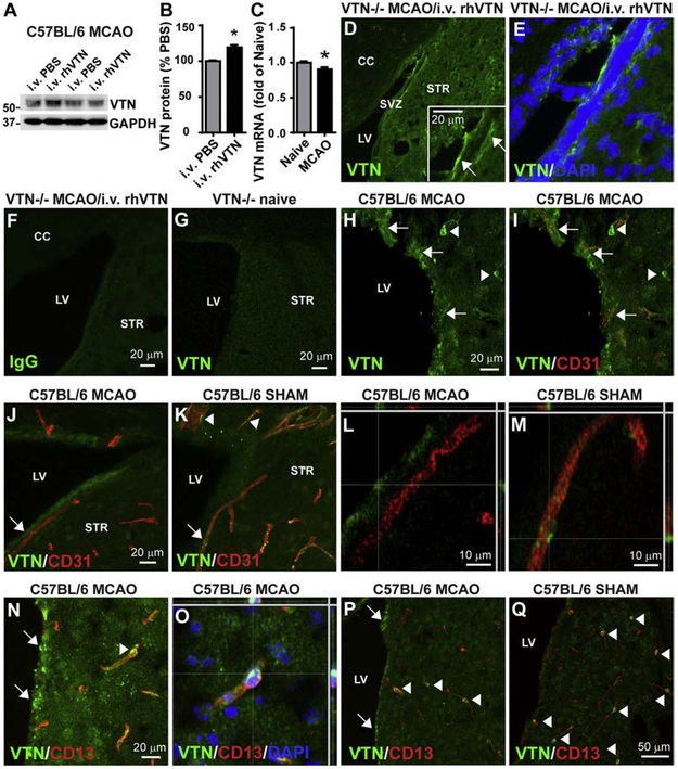
Figure 1. Blood VTN leaks into the SVZ following MCAO.
A) C57BL/6 mice injected i.v. with rhVTN at 24 h following MCAO show an increase of VTN protein in the SVZ 4 h later compared to PBS-injected mice. Different lanes in the representative western blot indicate SVZ samples from individual mice injected with PBS or rhVTN. B) Densitometry analysis from western blots of 3 independent gels and with n = 3 mice/group, * p<0.05 (T test). C) MCAO reduced VTN mRNA in the SVZ of C57BL/6 mice at 24 h. N = 3 and 5 mice. * p<0.05 (T-test). D,E) Deposits of i.v. injected rhVTN was detectable in the SVZ of VTN−/− mice by immunostaining. The insert in (D) is a higher magnification image and arrows indicate leaked VTN. No positive immunostaining was seen when the primary antibody was replaced by isotype IgG (F) or in the SVZ of naïve VTN−/− mice stained with VTN antibody (G). H) VTN deposits (green, indicated by arrows) were also detected in the SVZ of wildtype C57BL/6 mice 24 h following MCAO and were present around microvessels identified with the endothelial cell marker CD 31(red, I). Pericytes apposing blood vessels in the brain expressed VTN (arrowheads in K), as also shown by co-localization with the pericyte marker CD13 (red, N, arrowhead, shown in a confocal image in O). The number of VTN-positive pericytes was reduced at 24 h following MCAO (J,P) compared to sham-operated mice (K,Q). Arrowheads indicate VTN-positive (green) pericytes expressing CD13 (red). Arrows indicate leaked VTN in the SVZ. Scale bars are as indicated, CC, corpus callosum, LV, lateral ventricle, STR, striatum.

