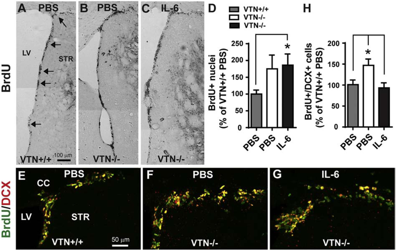
Figure 5. IL-6 blocks the female-specific VTN−/− phenotype of increased MCAO-induced neurogenesis.
Female VTN+/+ and VTN−/− mice were injected with PBS or IL-6 (100 ng) into the striatum next to the SVZ immediately prior to MCAO. BrdU were given the last 50 h before tissue collection at 14 days after MCAO. A-C) Representative images show that BrdU+ nuclei in the SVZ are more numerous in VTN−/− mice after PBS or IL-6 injection than in VTN+/+ mice after PBS injection. Arrows indicate the SVZ. LV, lateral ventricle, STR, striatum, scale bar = 100 μm. D) Unbiased stereological counting of BrdU+ nuclei in the SVZ were expressed as a percentage of PBS-injected VTN+/+ mice. This confirmed that VTN−/− mice have more proliferation after both PBS and IL-6 injection. N= 7, 3 and 6 mice. E-G) Representative images show that BrdU+/DCX+ cells in the dorsolateral SVZ of VTN+/+ and VTN−/− mice. The increase in VTN−/− mice seemed smaller after IL-6 compared to PBS injection. Scale bar = 50 μm. H) Quantification of BrdU+/DCX+ cells confirmed that VTN−/− mice have more neurogenesis than VTN+/+ mice, which was blocked by IL-6. N = 9, 3 and 6 mice. * p<0.05 (One-way ANOVA followed by post hoc Bonferroni multiple comparisons).

