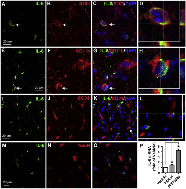
Figure 7. MCAO induces IL-6 protein expression in astrocytes and microglia/macrophages.
A 30 min of MCAO was performed in C57BL/6 mice and at 24 h IL-6 was detected in cells by double-immunofluorescent staining in the striatum close to SVZ. A-C) IL-6 (green) was colocalized in some astrocytes (arrow) identified with S100 staining (red), which is shown in a confocal image at higher magnification in (D). Nuclei were stained with DAPI (blue). E-H) IL-6 (green) was also present in some microglia/macrophages identified by CD11b (red). IL-6 was not present in endothelial cells (CD31, I-L) or neurons (NeuN, M-O). The areas indicated by arrows in C,G and K are shown with high magnification in D,H and L, respectively. Scale bar = 20 μm. P) Pharmacological inhibition of FAK in mouse primary astrocytes increases IL-6 expression. Mouse primary astrocyte cultures were incubated with vehicle (0.1% DMSO), FAK14 (10 μM) or PF573228 (10 μM) for 4 h and then IL-6 mRNA was measured by RT-qPCR. N = 3 independent experiments. * p<0.05 (paired one-way ANOVA followed by post hoc Bonferroni multiple comparisons).

