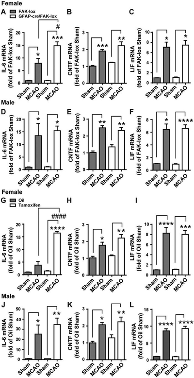
Figure 8. Astrocytic FAK deletion increases MCAO-induced IL-6 in females.

Female and male GFAP-cre/FAK-lox mice were treated for 5 days with tamoxifen to delete FAK in astrocytes. FAK-lox controls were treated the same. Two weeks after the last tamoxifen injection, they received a 30 min MCAO or a sham operation and the levels of cytokine mRNA in the SVZ were measured 24 later. Values were expressed as a fold change compared to sham-operated FAK-lox control mice. A) In females, MCAO caused an expected increase of IL-6 in FAK-lox controls (first two columns), and an even greater response in GFAP-cre/FAK-lox (last two columns). MCAO induced CNTF (B) and LIF (C) to the same extent in FAK-lox and GFAP-cre/FAK-lox females. In male mice, MCAO increased IL-6 (D), CNTF (E) and LIF (F) to the same extent in both FAK-lox and GFAP-cre/FAK-lox mice. N = 6, 9, 4 and 7 mice in females and N = 4, 5, 4 and 6 mice in males. To confirm that the effect of astrocyte FAK deletion were not due to lasting effects of tamoxifen, another set of GFAP-cre/FAK-lox mice was treated with oil or tamoxifen for 5 d and with an MCAO 14 d later. At 24 h following MCAO, SVZ mRNA expression was measured and calculated as a fold change from oil-treated sham operated mice within each sex. G) MCAO induced IL-6 was further increased with tamoxifen compared to oil control, which is consistent with data in (A). MCAO-induced CNTF (H) and LIF (I) were comparable between oil and tamoxifen treatment and within a similar range as seen in (B) and (C). In male mice, there were no differences of MCAO-induced IL-6 (J), CNTF (K) and LIF (L) between oil and tamoxifen-treated mice. N = 4, 4, 4 and 3 mice in females and N = 3, 4, 5 and 4 mice in males. * p<0.05, # p<0.05, ** p<0.01, *** p<0.001, **** p<0.0001 (Two-way ANOVA followed by post hoc Tukey multiple comparisons).
