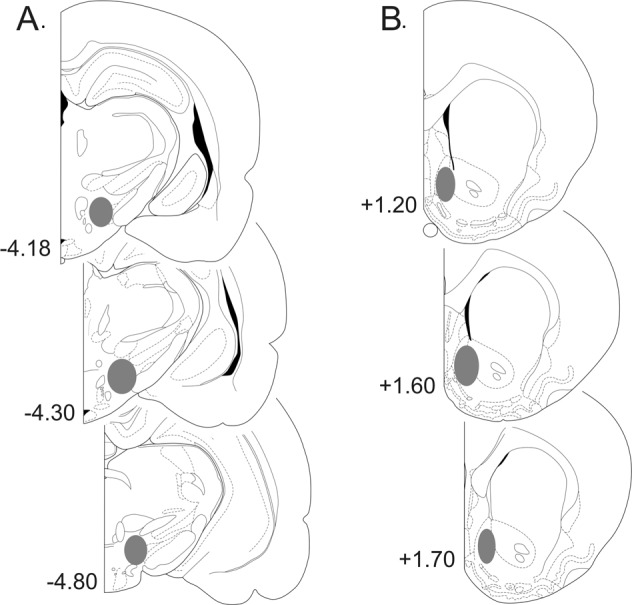
Fig. 2.

Representative coronal sections of the rat brain (adapted from the atlas of Paxinos and Watson29), with gray shaded areas indicating the placements of a stimulating electrodes in the medial forebrain bundle and b amperometric recording electrodes in the nucleus accumbens shell. Numbers correspond to mm from bregma
